Photo induced Force Microscopy (PiFM) combines the analytical power of infrared vibrational spectroscopy with the spatially resolved nano-topographical information of atomic force microscopy (AFM) creating an extraordinarily powerful technique for investigating the behaviour of surfaces in greater detail than has previously been possible. Whereas techniques such as SEM, EDX and TEM only identify the atoms themselves, the direct mapping of the surface chemistry with PiFM at a spatial resolution <10 nm includes all the advantages of vibrational spectroscopy such as isotopic labelling, identification of functional groups and molecular conformations and hence materials identification. Furthermore, there is a direct correlation between PiFM and standard lab FTIR spectra so that spectra can be directly compared with the IR databases that have been developed over the last 80 years.
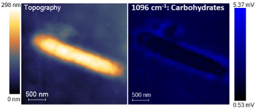
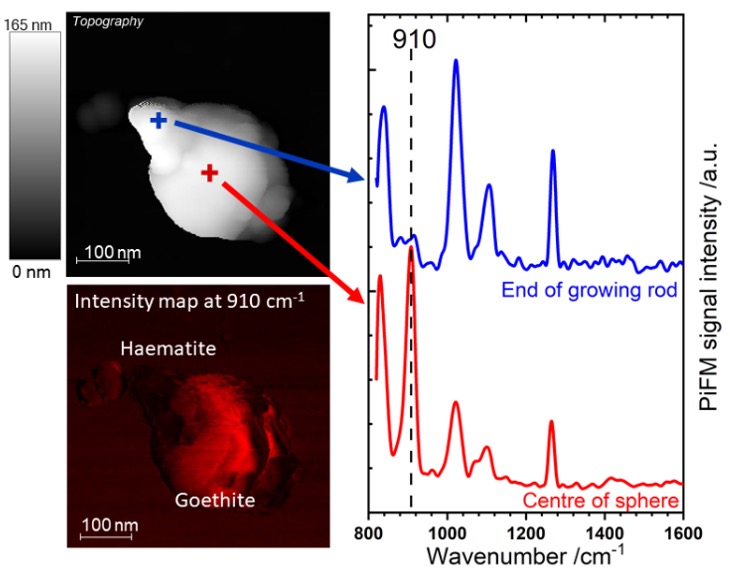
Samples are mounted in air as powders, crystals or films. A recent study of the growth of iron oxide nanorods for example, shows how the initial stages involve crystallisation as geothite an iron hydroxide, which subsequently recrystallises as haematite, Fig 1. Fig 2, shows the versatility of the PiFM with an image of an E. coli bacterium with the distribution of the vibrational band at 1096 cm-1 highlighting the carbohydrate shell that surrounds the bacterium.
Can PiFM help your work? Email daviespr@cardiff.ac.uk or daviesja21@cardiff.ac.uk and talk to us about what you want to do.